Seguem a diretrizes da Duke Sports Medicine Center.
Clinical Guideline for the Treatment of Adult Shoulder Pain
Clinical Guideline for the Treatment of Adult Knee Pain
Seguem a diretrizes da Duke Sports Medicine Center.
Clinical Guideline for the Treatment of Adult Shoulder Pain
Clinical Guideline for the Treatment of Adult Knee Pain
The skeletal muscle is a highly plastic tissue, namely, capable of adapting to adverse conditions. In nature, this intrinsic adaptive feature is crucial and ensures the survival of many species once a vast number of environmental impositions (e.g., contractile activity and mechanic overload) can be overcome by means of adaptive responses from the muscles. There are two main changes promoted by mechanical stimulation upon the skeletal muscle: (1) remodeling (namely, adaptation that induces changes in the muscle mass/volume); (2) alterations in the molecular engine of the muscle fiber, for example, phenotypical changes in myosin isoforms expression and important constituents of the skeletal muscle; (3) cellular and molecular adaptations leading to intracellular changes in specific aspects of muscle fiber, for example, mechanotransduction, expression of growth factors, and satellite cells activation. Taken together, these changes allow the muscle to adapt its contractile properties so that both power and strength are incremented.
With this information in mind, we invite investigators to submit relevant original papers as well as review articles that synthesize current knowledge or provide new data in the following issues. Potential topics inculde, but are not limited to:
Before submission authors should carefully read over the journal’s Author Guidelines, which are located at http://www.tswj.com/guidelines/. Prospective authors should submit an electronic copy of their complete manuscript through the journal Manuscript Tracking System athttp://mts.tswj.com/author/submit/physiology/musc/ according to the following timetable:
| Manuscript Due | Friday, 30 November 2012 |
| Final Decision Date | Friday, 28 December 2012 |
| Publication Date | Friday, 8 Fabruary 2013 |
Novartis Institutes for Biomedical Research, 100 Technology Square, Cambridge, MA 02139, USA.
Skeletal muscle mass is regulated by activity, metabolism, and the availability of nutrients. During muscle atrophy, MNK2 expression increases. We found that MNK2 (mitogen-activated protein kinase-interacting kinase 2), but not MNK1, inhibited proteins involved in promoting protein synthesis, including eukaryotic translation initiation factor 4G (eIF4G) and mammalian target of rapamycin (mTOR). Phosphorylation at serine 1108 (Ser¹¹⁰⁸) of eIF4G, which is associated with enhanced protein translation, is promoted by insulin-like growth factor 1 and inhibited by rapamycin or starvation, suggesting that phosphorylation of this residue is regulated by mTOR. In cultured myotubes, small interfering RNA (siRNA) knockdown of MNK2 increased eIF4G Ser¹¹⁰⁸ phosphorylation and overcame rapamycin’s inhibitory effect on this phosphorylation event. Phosphorylation of Ser¹¹⁰⁸ in eIF4G, in gastrocnemius muscle, was increased in mice lacking MNK2, but not those lacking MNK1, and this increased phosphorylation was maintained in MNK2-null animals under atrophy conditions and upon starvation. Conversely, overexpression of MNK2 decreased eIF4G Ser¹¹⁰⁸ phosphorylation. An siRNA screen revealed that serine-arginine-rich protein kinases linked increased MNK2 activity to decreased eIF4G phosphorylation. In addition, we found that MNK2 interacted with mTOR and inhibited phosphorylation of the mTOR target, the ribosomal kinase p70S6K (70-kD ribosomal protein S6 kinase), through a mechanism independent of the kinase activity of MNK2. These data indicate that MNK2 plays a unique role, not shared by its closest paralog MNK1, in limiting protein translation through its negative effect on eIF4G Ser¹¹⁰⁸ phosphorylation and p70S6K activation.
O Professor Marc Francaux lançou ontem no Twitter:
 O Congresso Internacional de Bioquímica do Exercício acontece trienalmente e sua próxima edição será em São Paulo!
Site da edição deste ano em Estocolmo: http://www.ibec2012.org/
Resumo do trabalho:
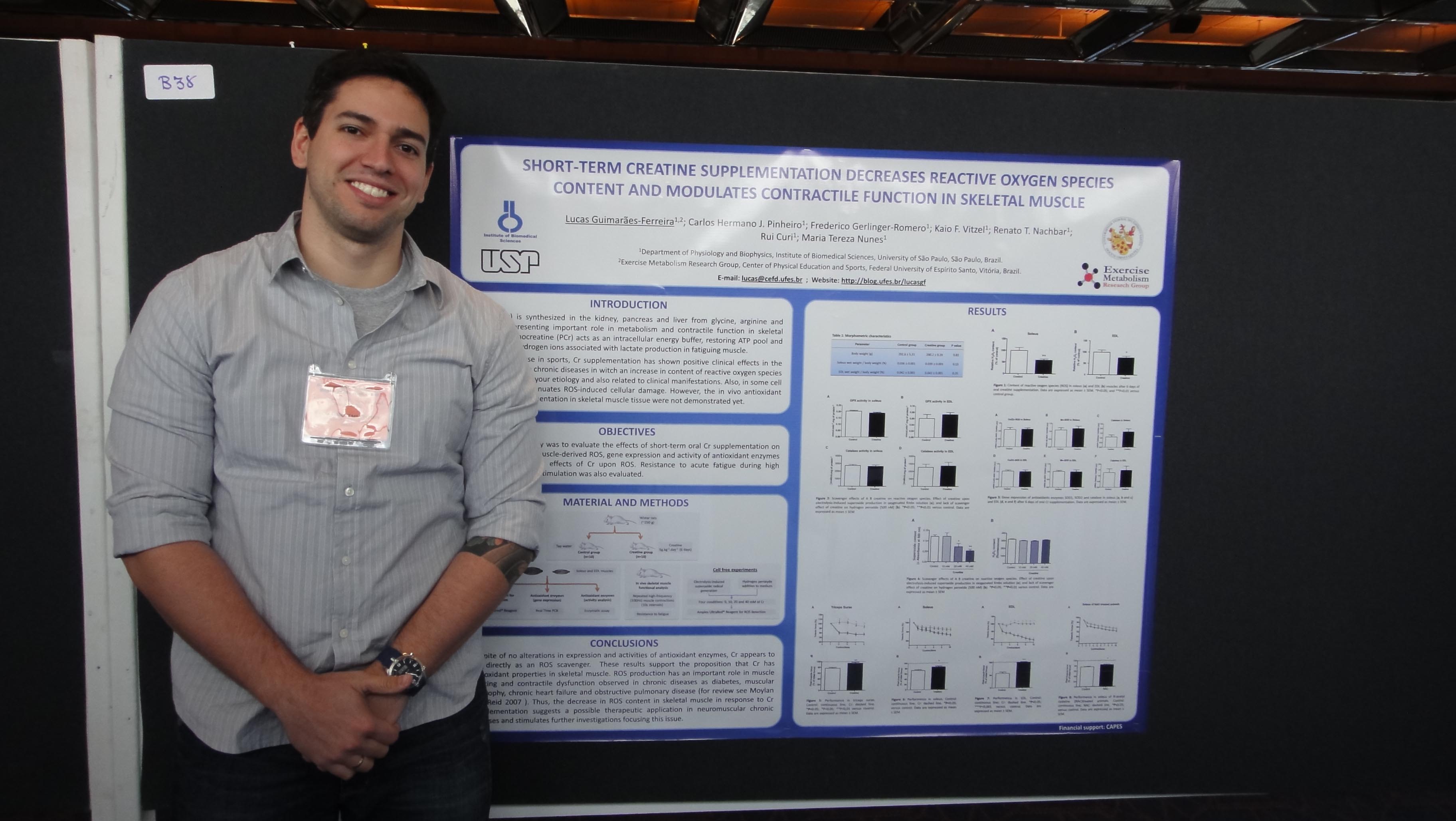
Short-term creatine supplementation decrease reactive oxygen species content and modulates contractile function in skeletal muscle
Lucas Guimarães-Ferreira; Carlos Hermano J. Pinhero; Frederico Gerlinger-Romero; Kaio F. Vitzel; Renato T. Nachbar; Rui Curi; Maria Tereza Nunes
Department of Physiology and Biophysics, Institute of Biomedical Sciences, University of São Paulo, São Paulo/SP – Brazil.
Exercise Metabolism Research Group, Center of Physical Education and Sports, Federal University of Espirito Santo, Vitória/ES – Brazil.
Email: lucas@cefd.ufes.br.
The effect of short-term creatine supplementation (CrS) upon contractile function and content of reactive oxygen species (ROS) in skeletal muscle was investigated. Wistar rats were supplemented with creatine (Cr; 5 g/kg BW) by gavage for 6 days. Muscle contractions were evoked in triceps surae, soleus and extensor digitorum longus (EDL) in different experiments by direct electrical stimulation (ES) of sciatic nerve in vivo. Resistance to acute fatigue of rat skeletal muscle were investigated. Soleus and EDL muscles were incubated for evaluation of ROS content using Amplex-UltraRed reagent. The analysis of expression and activity of antioxidant enzymes were performed. Direct scavenger action of Cr on superoxide radical and hydrogen peroxide was also investigated. CrS improves resistance to acute fatigue during high frequency ES, evaluated by fall in force production during sucessives isometric contractions in triceps surae (control: 23%; Cr: 6.5%), soleus (control: 27.1%; Cr: 14.9%)and EDL (control: 43.2%; Cr: 0.1%) muscles. ROS content was also decreased in soleus (by 41%) and EDL (by 33.7%) muscles from Cr-supplemented rats. CrS did not change expression and activity of antioxidant enzymes. In cell-free experiments, Cr showed a scavenger action on superoxide radical, but not in hydrogen peroxide. These results indicate that CrS decreases ROS content in skeletal muscle possibly due to a direct action of Cr on superoxide radical. This action may account to CrS effects on contractile function, which were similar to those observed when N-acetyl-cysteine, a potent antioxidant, is administered prior to ES.
The Åstrand Laboratory, Swedish School of Sport and Health Sciences, Stockholm, Sweden.
Resistance exercise and amino acids are two major factors that influence muscle protein turnover. Here, we examined the effects of resistance exercise and branched-chain amino acids (BCAA), individually and in combination, on the expression of anabolic and catabolic genes in human skeletal muscle. Seven subjects performed two sessions of unilateral leg press exercise with randomized supplementation with BCAA or flavored water. Biopsies were collected from the vastus lateralis muscle of both the resting and exercising legs before and repeatedly after exercise to determine levels of mRNA, protein phosphorylation, and amino acid concentrations. Intake of BCAA reduced (P < 0.05) MAFbx mRNA by 30 and 50% in the resting and exercising legs, respectively. The level of MuRF-1 mRNA was elevated (P < 0.05) in the exercising leg two- and threefold under the placebo and BCAA conditions, respectively, whereas MuRF-1 total protein increased by 20% (P < 0.05) only in the placebo condition. Phosphorylation of p70(S6k) increased to a larger extent (∼2-fold; P < 0.05) in the early recovery period with BCAA supplementation, whereas the expression of genes regulating mTOR activity was not influenced by BCAA. Muscle levels of phenylalanine and tyrosine were reduced (13-17%) throughout recovery (P < 0.05) in the placebo condition and to a greater extent (32-43%; P < 0.05) following BCAA supplementation in both resting and exercising muscle. In conclusion, BCAA ingestion reduced MAFbx mRNA and prevented the exercise-induced increase in MuRF-1 total protein in both resting and exercising leg. Further-more, resistance exercise differently influenced MAFbx and MuRF-1 mRNA expression, suggesting both common and divergent regulation of these two ubiquitin ligases.
Este vídeo mostra: microRNA formation and function
Department of Physiology and Biophysics, Institute of Biomedical Sciences, University of São Paulo, ICB-I, Cidade Universitária, Av. Prof. Lineu Prestes, 1524, Butantã, São Paulo, SP, 05508-900, Brazil, lucas@cefd.ufes.br.
The effect of short-term creatine (Cr) supplementation upon content of skeletal muscle-derived-reactive oxygen species (ROS) was investigated. Wistar rats were supplemented with Cr (5 g/kg BW) or vehicle, by gavage, for 6 days. Soleus and extensor digitorum longus (EDL) muscles were removed and incubated for evaluation of ROS content using Amplex-UltraRed reagent. The analysis of expression and activity of antioxidant enzymes (superoxide dismutase 1 and 2, catalase and glutathione peroxidase) were performed. Direct scavenger action of Cr on superoxide radical and hydrogen peroxide was also investigated. Short-term Cr supplementation attenuated ROS content in both soleus and EDL muscles (by 41 and 33.7%, respectively). Cr supplementation did not change expression and activity of antioxidant enzymes. Basal TBARS content was not altered by Cr supplementation. In cell-free experiments, Cr showed a scavenger effect on superoxide radical in concentrations of 20 and 40 mM, but not on hydrogen peroxide. These results indicate that Cr supplementation decreases ROS content in skeletal muscle possibly due to a direct action of Cr molecule on superoxide radical.
Nosso estudo recentemente publicado no European Journal of Applied Physiology traz novos dados que apontam para possíveis efeitos terapêuticos da suplementação com creatina, especialmente em doenças neuromusculares.
Doenças como diabetes, distrofia muscular, DPOC, insuficiência cardíaca e outras têm em comum uma produção exacerbada de espécies reativas de oxigênio (ROS). Em nosso estudo, mostramos que após 6 dias de suplementação com creatina monohidratada, o conteúdo de ROS no músculo esquelético foi significativamente menor nos músculos sóleo e EDL.

A expressão gênica e atividade das enzimas antioxidantes não foi alterada. Entretanto, em experimentos cell free demonstramos que a creatina pode atuar como scavenger contra o ânion superóxido, mas não o peróxido de hidrogênio.

Concluindo, nossos dados suportam a ação antioxidante da creatina, demonstrando pela primeira vez uma redução de ROS no músculo esquelético in vivo. Esta ação parece envolver uma ação direta da molécula de creatina sobre ROS.